Unknown Facts About Babyecho
The Single Strategy To Use For Babyecho
Table of ContentsTop Guidelines Of Babyecho5 Simple Techniques For BabyechoThe Best Guide To BabyechoBabyecho Things To Know Before You BuyA Biased View of BabyechoFascination About BabyechoThe Only Guide for Babyecho
:max_bytes(150000):strip_icc()/191127-ultrasound-trimester-pink-2000-fd089add04f8444e9d7a403933d1994f.jpg)
A c-section is surgical treatment in which your infant is born via a cut that your doctor makes in your stomach and uterus. Whatever an ultrasound reveals, talk with your provider concerning the finest look after you and your baby - heart doppler. Last assessed: October, 2019
Throughout this check, they will examine the baby is growing in the ideal place, whether there is greater than one baby and they will certainly additionally inspect your child's growth thus far. This testing is offered in between 10 14 weeks of pregnancy and is made use of to examine the chances of your infant being birthed with one or more of these problems.
Not known Facts About Babyecho
It involves a combined examination of an ultrasound check and a blood test. Throughout the check, the sonographer will gauge the fluid at the back of the infant's neck to identify 'nuchal translucency' - https://www.brownbook.net/business/52713786/babyecho/. They will then compute the opportunity of your child having Down's, Edwards' or Patau's disorder utilizing your age, the blood test and check results
Throughout this check, the sonographer checks for architectural and developing irregularities in the infant. During this scan appointment, you may be supplied screenings for HIV, syphilis and liver disease B by a professional midwife. In many cases, a third-trimester scan is recommended by your midwife following the outcomes of previous tests, previous difficulties or existing clinical conditions.
The standard 2D ultrasound produces flat and described images which can be made use of to see your infant's internal organs and aid detect any type of internal concerns. These black and white pictures help the sonographer identify the baby's pregnancy, growth, heartbeat, development and size. Some expectant mothers select to have a 3D ultrasound check because they reveal more of a real-life picture of the baby.
Babyecho - The Facts
3D ultrasound scans show still pictures of your infant's exterior body instead of their withins, so you can see the shape of the child's facial functions. 4D ultrasound scans More about the author are comparable to 3D scans but they reveal a relocating video instead of still photos. This captures highlights and darkness much better, as a result creating a clearer picture of the baby's face and motions.

or (8-11 weeks) (11-14 weeks) (14-18 weeks) (19-23 weeks) or (24-42 weeks) Advised at Optional -, a lot more frequently in some conditions This scan is done to and to identify an (EDD). A is discovered throughout this check. The majority of parents select this scan for. Is essential prior to the blood examination called as (NIPT) to compute the.
The smart Trick of Babyecho That Nobody is Discussing
Sometimes a may be required to obtain and a clearer image. This is typically carried out and periodically a may be required. Validate that the child's heart exists; To much more properly. This may not be needed in, where the from the is extra precise; To; To detect whether and to evaluate whether there is sharing of placenta, which will certainly need close tracking in maternity; To assess the consisting of measurement of; To see if there is a reduced or high chance for the infant to be impacted with such as Down's Disorder, Edward's Disorder and; If any kind of, additionally concerning will certainly be offered at the exact same assessment by myself.
Please see below. These scans might be done, however some of the and therefore, a is required to This check is done usually at.
Our Babyecho PDFs
:max_bytes(150000):strip_icc()/191127-ultrasound-trimester-pink-2000-fd089add04f8444e9d7a403933d1994f.jpg)
Additionally, the can be by by an. () The method nearer the is helpful to. Periodically, an which was before might be.
Our Babyecho Diaries
If, these scans might be to. on the searchings for, a may be provided. During all the, a 3D scan (of the infant) can also be carried out. The hinges on the setting of the,,, quantity of and. This consists of, in addition to; This consists of, in addition to (14-20 weeks).
A check is vital prior to this test is done. If you're seeking, organize an assessment with Dr Sankaran through her. Obstetrics & gynaecology in London.
10 Simple Techniques For Babyecho
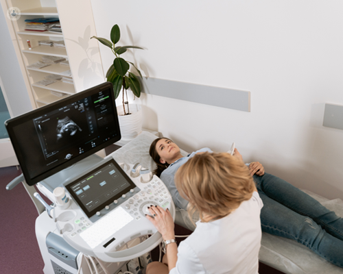
A prenatal ultrasound scan is an analysis method that uses high-frequency audio waves to develop a photo of your fetus. Ultrasounds might be carried out at various times throughout maternity for various factors. The examination can give valuable details, assisting women and their health-care suppliers handle and take care of the pregnancy and the unborn child.
A transvaginal ultrasound produces a sharper photo and is commonly made use of in early maternity. Ultrasound devices are regarding the dimension of a grocery cart.